Treatments

Coronary Angiography: A Vital Diagnostic Tool for Heart Health

Coronary angiography is a specialized diagnostic procedure that provides detailed images of the heart's blood vessels, helping cardiologists evaluate and manage heart diseases. It is considered the gold standard for identifying blockages or abnormalities in the coronary arteries.

What is Coronary Angiography?

Coronary angiography is a minimally invasive procedure that uses X-ray imaging to visualize the coronary arteries. A contrast dye is injected into the arteries through a thin catheter, highlighting blood flow and revealing any blockages or narrowing that may impede the heart’s function.

Why is Coronary Angiography Performed?

This test is often recommended for individuals experiencing symptoms of heart disease or those with risk factors requiring further investigation. Indications include:

  • Persistent chest pain (angina).
  • Shortness of breath.
  • Abnormal results from other tests like stress tests or echocardiograms.
  • Diagnosing coronary artery disease (CAD).
  • Assessing heart health before surgery.

How is the Procedure Done?

1. Preparation:

  • The patient is given local anesthesia at the insertion site (usually the wrist or groin).
  • Vital signs are monitored throughout the procedure.

2. Catheter Insertion:

  • TA thin catheter is guided through a blood vessel to the coronary arteries.

3. Contrast Dye Injection:

  • A special dye is injected, making the coronary arteries visible on X-ray images.

4. Imaging:

  • X-ray imaging captures detailed views of the coronary arteries, highlighting any blockages or abnormalities.

The procedure typically lasts 30-60 minutes, and most patients can return home the same day.

Benefits of Coronary Angiography

  • Accurate Diagnosis: Provides precise information about the location and severity of blockages.
  • Guided Treatment: Helps plan further interventions like angioplasty, stenting, or bypass surgery.
  • Early Detection: Identifies potential problems before they lead to severe complications.

Is Coronary Angiography Safe?

Coronary angiography is a safe and commonly performed procedure, with a low risk of complications. Any risks, such as minor bleeding at the catheter insertion site or allergic reactions to the dye, are carefully managed by the medical team.

What to Expect After the Procedure

  • Patients are advised to rest for a few hours and avoid strenuous activities for a day or two.
  • Follow-up care is essential to discuss results and plan further treatment if needed.

Why Choose Our Expertise in Coronary Angiography?

  • Experienced Cardiologist: Performed by a skilled interventional cardiologist with expertise in complex procedures.
  • Advanced Technology: State-of-the-art imaging equipment ensures accurate and detailed results.
  • Patient-Centered Care: Comprehensive pre- and post-procedure guidance to ensure a smooth and comfortable experience.

Take control of your heart health today. Schedule your coronary angiography with our expert Dr. Akshat Jain and gain clarity on your cardiovascular well-being.

Coronary Angioplasty: Restoring Blood Flow, Reviving Heart Health

Coronary angioplasty, also known as percutaneous coronary intervention (PCI), is a minimally invasive procedure used to open clogged or narrowed coronary arteries, restoring normal blood flow to the heart. It is a life-saving treatment for individuals experiencing chest pain (angina) or heart attacks caused by blockages.

What is Coronary Angioplasty?

Coronary angioplasty involves the use of a small balloon and a stent to widen blocked or narrowed arteries. By restoring blood flow, the procedure reduces symptoms like chest pain and prevents serious complications such as heart attacks.

When is Coronary Angioplasty Needed?

This procedure is recommended for patients with:

  • Persistent angina (Chest Pain) that doesn’t respond to medications.
  • Acute myocardial infarction (heart attack).
  • Severe narrowing or blockages detected during coronary angiography.
  • A high risk of coronary artery disease complications.

How is Coronary Angioplasty Performed?

1. Preparation:

  • The patient is given local anesthesia and, if needed, mild sedation.
  • A catheter is inserted through an artery, usually in the wrist or groin.

2. Balloon Inflation:

  • A thin, flexible tube with a deflated balloon at its tip is guided to the blockage.
  • The balloon is inflated to compress the plaque against the artery walls, widening the vessel.

3. Stent Placement:

  • A stent (a small, mesh-like metal tube) is often placed to keep the artery open and prevent re-narrowing.

The procedure typically takes 30 minutes to 2 hours, depending on the complexity of the blockages.

Types of Stents Used in Angioplasty

  • Bare-Metal Stents (BMS): Provide structural support to the artery.
  • Drug-Eluting Stents (DES): Coated with medication to reduce the risk of re-narrowing.
  • Bioabsorbable Stents: Dissolve over time, leaving the artery unobstructed.

Benefits of Coronary Angioplasty

  • Immediate Symptom Relief: Reduces or eliminates chest pain and breathlessness.
  • Minimally Invasive: Quicker recovery compared to open-heart surgery.
  • Life-Saving in Emergencies: Quickly restores blood flow during heart attacks.
  • Improves Quality of Life: Enhances physical activity levels and overall well-being.

Recovery After Coronary Angioplasty

  • Most patients can return to normal activities within a few days.
  • Lifestyle changes, such as a healthy diet, exercise, and quitting smoking, are crucial for long-term success.
  • Medications like blood thinners may be prescribed to prevent clot formation in the stent.

Why Choose Our Expertise in Coronary Angioplasty?

  • Expert Care: Performed by an experienced interventional cardiologist with a proven track record in handling complex cases.
  • Advanced Facilities: Equipped with state-of-the-art cath labs and imaging technology.
  • Comprehensive Support: From initial consultation to follow-up care, we provide patient-centered treatment tailored to your needs.

Take Charge of Your Heart Health Today

If you’re experiencing symptoms of heart disease, don’t wait. Book a consultation with our cardiologist Dr. Akshat Jain and explore how coronary angioplasty can transform your life.

Coronary Angioplasty with Drug-Coated Balloon: A Stent-Free Solution for Heart Health

Coronary angioplasty with a drug-coated balloon (DCB) is an innovative, stent-free procedure that effectively treats narrowed or blocked coronary arteries. This cutting-edge approach offers an alternative to traditional stenting, particularly in specific cases where long-term stent placement may not be ideal.

What is Coronary Angioplasty with Drug-Coated Balloon?

This procedure involves the use of a specialized balloon coated with medication to open blocked arteries and prevent re-narrowing. Unlike traditional angioplasty, a stent is not implanted, leaving the artery free of any permanent foreign object. The drug released from the balloon helps inhibit cell proliferation, reducing the risk of restenosis (re-narrowing) after the procedure.

Who Can Benefit from This Procedure?

Drug-coated balloon angioplasty is especially beneficial in the following scenarios:

  • Small Arteries: Where placing a stent may be challenging.
  • In-Stent Restenosis: Treating blockages that recur inside previously placed stents.
  • Patients at High Bleeding Risk: Reduces the need for long-term blood-thinning medications.
  • Bifurcation Lesions: Ideal for treating branch-like structures of coronary arteries.

How is the Procedure Performed?

The procedure is performed using the following steps:

1. Preparation:

  • Local anesthesia is administered, and a catheter is inserted through the wrist or groin.

2. Balloon Inflation:

  • A drug-coated balloon is guided to the blockage.
  • The balloon is inflated, compressing the plaque and delivering medication to the artery wall.

3. Deflation and Removal:

  • Once the medication is delivered, the balloon is deflated and removed, leaving the artery open without a stent.

The procedure is typically completed in less than an hour and is minimally invasive.

Advantages of Drug-Coated Balloon Angioplasty

Drug-coated balloon angioplasty offers several advantages over traditional stenting:

  • Stent-Free Treatment: Eliminates the need for permanent implants.
  • Reduced Risk of Complications: Lowers the risk of stent thrombosis and long-term bleeding.
  • Minimally Invasive: Faster recovery and minimal discomfort.
  • Preserves Future Treatment Options: Keeps arteries clear for potential future interventions.

Recovery and Aftercare

  • Patients usually return home the same day or within 24-48 hours.
  • Lifestyle modifications, such as a healthy diet and regular exercise, are essential for long-term success.
  • Follow-up care includes regular check-ups and medications to ensure optimal artery health.

Why Choose Our Expertise in Drug-Coated Balloon Angioplasty?

  • Specialized Expertise: Performed by an experienced interventional cardiologist skilled in advanced, stent-free techniques.
  • State-of-the-Art Technology: Our cath lab is equipped with the latest tools for precision and safety.
  • Comprehensive Care: From initial evaluation to post-procedure follow-ups, we provide seamless, patient-focused care.

Is Drug-Coated Balloon Angioplasty Right for You?

If you’ve been diagnosed with coronary artery disease or have recurring blockages, this innovative procedure could be the ideal solution for you.

Contact Dr. Akshat Jain today to schedule a consultation and learn how this stent-free treatment can improve your heart health.

Advanced Angioplasty Techniques for Complex Lesion Preparation: Cutting Balloon, Laser Ablation, Rotational Atherectomy, Orbital Atherectomy, and Intravascular Lithotripsy (IVL)

Advanced angioplasty techniques are critical tools in the management of complex coronary artery disease (CAD), particularly when dealing with challenging lesions that traditional methods cannot treat effectively. These techniques are designed to prepare difficult-to-treat coronary blockages for stent placement or balloon angioplasty, ensuring optimal outcomes for patients. For patients under the care of Dr. Akshat Jain, advanced interventions like Cutting Balloon, Laser Ablation, Rotational Atherectomy, Orbital Atherectomy, and Intravascular Lithotripsy (IVL) provide cutting-edge solutions to coronary artery challenges, restoring normal blood flow with precision and safety.

Cutting Balloon: Precision for Resistant Lesions

The Cutting Balloon is a specialized balloon catheter equipped with micro-incisions or blades on its surface. It is particularly useful for calcified or fibrotic lesions that are resistant to conventional balloon angioplasty. When inflated, the blades gently score the plaque, allowing for better vessel dilation and stent placement.

Key Benefits of Cutting Balloon:

  • Targeted Plaque Modification: Perfect for treating tough, calcified lesions that do not respond well to traditional balloons.
  • Reduced Risk of Vessel Injury: The controlled cutting action minimizes the risk of dissection and damage to the vessel wall.
  • Improved Stent Expansion: Enhances stent deployment by ensuring optimal vessel size for stent placement.

Laser Ablation: High-Energy Light for Challenging Lesions

Laser Ablation uses a catheter equipped with a laser fiber to emit high-energy light that vaporizes and removes hard plaques inside coronary arteries. This technique is especially effective for severe calcifications and fibrotic plaques, which are often difficult to treat using standard angioplasty.

Key Benefits of Laser Ablation:

  • Effective for Dense Plaque: Ideal for heavily calcified or fibrotic lesions that are resistant to other interventions.
  • Minimal Risk to Vessel Wall: Laser energy precisely targets the plaque, reducing the risk of vessel injury.
  • Effective for Chronic Total Occlusions (CTOs): Useful for opening blocked arteries that may not be amenable to other treatments.

Rotational Atherectomy: Precision Debulking for Calcified Lesions

Rotational Atherectomy (also known as Rotablation) involves using a high-speed, diamond-coated burr to rotate within the artery, grinding up calcified plaques. This technique is particularly beneficial for severely calcified lesions that resist conventional balloon angioplasty. The rotating burr breaks down the plaque into small particles, which are then washed away by blood flow.

Key Benefits of Rotational Atherectomy:

  • Effective for Calcified Lesions: Ideal for treating hard, calcified blockages that are difficult to dilate with standard balloons.
  • Helps in Stent Preparation: Facilitates stent deployment by preparing the artery and creating space for optimal stent placement.
  • Precision Debulking: Helps remove large amounts of plaque in a controlled manner, improving artery expansion.

Orbital Atherectomy: Advanced Plaque Modification

Orbital Atherectomy is a unique technique that uses a diamond-coated crown to orbit around the lesion at high speeds, safely shaving off calcified plaque. This device rotates in an orbital motion, which helps dislodge plaque without damaging the arterial wall. Orbital atherectomy is ideal for severely calcified lesions and is often used when rotational atherectomy may be too aggressive.

Key Benefits of Orbital Atherectomy:

  • Safe for Severe Calcification: Perfect for removing hard, calcified plaque without harming the vessel wall.
  • Precision and Control: Orbital motion allows for targeted plaque removal, reducing the risk of injury.
  • Effective for Complex Lesions: Often used in cases of chronic total occlusions (CTOs) or heavily calcified lesions that require a more delicate approach.

Intravascular Lithotripsy (IVL): Shockwave Technology for Calcified Lesions

Intravascular Lithotripsy (IVL) is an innovative technique that uses shockwave technology to fracture calcified plaques within coronary arteries. A balloon catheter is placed at the site of the lesion, and shockwaves are emitted, creating microfractures in the calcified plaque, making it easier to dilate the vessel and prepare it for stenting. IVL is especially useful in severely calcified lesions that resist conventional balloon dilation.

Key Benefits of Intravascular Lithotripsy (IVL):

  • Non-Thermal and Non-Mechanical: Uses shockwaves rather than heat or mechanical force, reducing the risk of damage to the vessel wall.
  • Effective for Heavily Calcified Lesions: Particularly useful for treating tough, calcified plaques that traditional methods may not address effectively.
  • Minimal Risk of Dissection: Shockwaves are precisely controlled to ensure minimal vessel injury while breaking up plaque.
  • Optimizes Stent Deployment: IVL prepares the vessel, allowing for better stent expansion and apposition, improving long-term results.

Why Choose Dr. Akshat Jain for Advanced Angioplasty Techniques?

  • Expert in Complex Lesion Management: Dr. Akshat Jain brings years of experience in handling complex coronary lesions, utilizing these advanced techniques to ensure the best patient outcomes.
  • State-of-the-Art Equipment: Our Hospital provides with the latest technology for performing Cutting Balloon, Laser Ablation, Rotational Atherectomy, Orbital Atherectomy, and Intravascular Lithotripsy (IVL), ensuring precision and safety throughout the procedure.
  • Personalized Treatment Plans: Dr. Jain tailors each approach to the unique needs of the patient, selecting the most suitable technique to ensure successful treatment of complex lesions.
  • Comprehensive Care: From diagnosis to recovery, Dr. Jain offers personalized care, guiding patients through every step of their treatment journey.

Restore your heart health with advanced angioplasty techniques. If you or a loved one is dealing with complex coronary artery disease, advanced interventions like Cutting Balloon Angioplasty, Laser Ablation, Rotational Atherectomy, Orbital Atherectomy, and Intravascular Lithotripsy (IVL) can provide effective solutions to restore normal blood flow and improve quality of life. Contact Dr. Akshat Jain today to explore your treatment options and take the next step towards a healthier heart.

Intracoronary Imaging and Physiology Studies: Advanced Techniques for Precision in Cardiovascular Care

Intracoronary Imaging and Physiology studies are essential tools in modern interventional cardiology, providing invaluable insights into coronary artery disease (CAD) that go beyond traditional angiography. These advanced techniques help interventional cardiologists, like Dr. Akshat Jain, precisely assess the condition of coronary arteries and make informed decisions for treatment. Techniques like Intravascular Ultrasound (IVUS), Optical Coherence Tomography (OCT), Fractional Flow Reserve (FFR), and Resting Fractional Flow Reserve (RFR) provide detailed images and physiological data, enhancing patient outcomes through personalized treatment strategies.

Intravascular Ultrasound (IVUS): Detailed 3D Visualization of Coronary Arteries

IVUS is a powerful imaging tool that uses high-frequency sound waves to create detailed, cross-sectional images of coronary arteries. By providing a 360-degree view of the vessel walls, IVUS enables cardiologists to evaluate the severity of atherosclerotic plaques, the presence of thrombus (blood clots), and the structure of the vessel. It plays a crucial role in guiding coronary interventions such as stent placement.

Key Benefits of IVUS:

  • Accurate Vessel Sizing: Ensures optimal stent size and placement to minimize complications.
  • Assessment of Plaque Burden: Helps determine the degree of plaque accumulation, which is critical in treatment decisions.
  • Stent Optimization: Provides real-time data to optimize stent deployment and assess post-procedural results.

Optical Coherence Tomography (OCT): High-Resolution Imaging for Coronary Artery Assessment

OCT is a cutting-edge optical imaging technique that uses infrared light to capture high-resolution images of coronary arteries. With its ability to produce micrometer-level images, OCT offers superior resolution compared to IVUS, allowing for precise assessment of the arterial walls, plaque composition, and stent placement.

Key Benefits of OCT:

  • Superior Resolution: Ideal for visualizing fine details of the coronary vessel, including the intima-media layer and plaque structures.
  • Stent and Device Assessment: Enables detailed evaluation of stent apposition and expansion, crucial for minimizing restenosis.
  • Detection of Vulnerable Plaques: Identifies thin-cap fibroatheromas (TCFA) or other high-risk plaque characteristics, guiding early intervention strategies.

Fractional Flow Reserve (FFR): Measuring Coronary Artery Function

FFR is a physiological index that measures the pressure difference across a coronary stenosis during cardiac catheterization. It provides a functional assessment of coronary lesions by evaluating the extent to which a blockage restricts blood flow to the heart muscle. This information is critical in determining whether a coronary lesion requires intervention, such as angioplasty or stenting.

Key Benefits of FFR:

  • Functional Assessment: Helps determine the clinical significance of coronary lesions, guiding treatment decisions.
  • Avoids Unnecessary Interventions: By measuring the physiological impact of stenosis, FFR helps avoid unnecessary procedures, ensuring that only lesions that truly impair blood flow are treated.
  • Improved Patient Outcomes: FFR-guided interventions have been shown to lead to better long-term outcomes compared to angiography alone.

Resting Fractional Flow Reserve (RFR): A Non-Invasive Alternative to FFR

RFR is a novel, non-invasive method to assess coronary artery function without the need for adenosine, which is typically used in FFR testing to induce maximal hyperemia (increased blood flow). Using the pressure measurements obtained during routine coronary angiography, RFR provides a resting index of coronary flow reserve, offering a less invasive and more efficient alternative for lesion assessment.

Key Benefits of RFR:

  • Non-Invasive: Eliminates the need for additional pharmacological agents (like adenosine) required in traditional FFR.
  • Efficient Assessment: Can be quickly measured during routine coronary angiography, streamlining the diagnostic process.
  • Accurate and Reliable: Provides similar diagnostic information to FFR, helping determine the need for intervention.

Why Choose Dr. Akshat Jain for Intracoronary Imaging and Physiology Studies?

  • Expert Interventional Cardiologist: Dr. Akshat Jain is highly skilled in utilizing advanced imaging and physiology tools to provide comprehensive care. With expertise in interpreting IVUS, OCT, FFR, and RFR, Dr. Jain ensures precise, data-driven decisions for coronary interventions.
  • State-of-the-Art Technology: Our Hospital is equipped with the latest diagnostic tools and imaging systems, ensuring high-resolution images and accurate physiological data for optimal treatment outcomes.
  • Personalized Treatment Plans: By leveraging these advanced techniques, Dr. Jain tailors each treatment to the unique needs of the patient, ensuring the most effective and least invasive approach for managing coronary artery disease.

Take control of your heart health today. Advanced intracoronary imaging and physiology studies like IVUS, OCT, FFR, and RFR offer critical insights into coronary artery disease, enabling precise diagnosis and treatment planning. If you or a loved one is experiencing symptoms of heart disease, contact Dr. Akshat Jain to explore how these advanced diagnostic techniques can improve care and outcomes.

Conventional Single and Dual Chamber Pacemaker Implantation: Restoring Heart Rhythm

Pacemakers are life-saving devices designed to regulate abnormal heart rhythms, ensuring your heart beats efficiently and maintains adequate blood flow throughout your body. Conventional single and dual-chamber pacemakers are among the most commonly used devices to manage bradycardia (slow heart rate) and other rhythm disorders.

What is a Pacemaker?

A pacemaker is a small, battery-powered device implanted under the skin, usually near the collarbone. It sends electrical impulses to the heart to maintain a normal rhythm and rate.

  • Single-Chamber Pacemaker: Stimulates one chamber of the heart, either the right atrium or the right ventricle.
  • Dual-Chamber Pacemaker: Sends signals to both the right atrium and the right ventricle, coordinating their activity to mimic the heart's natural rhythm.

Who Needs a Pacemaker?

Pacemaker implantation is recommended for patients with:

  • Bradycardia: Abnormally slow heart rate.
  • Heart Block: Disrupted electrical signals between the atria and ventricles.
  • Atrial Fibrillation with Slow Ventricular Response: To maintain an adequate ventricular rate.
  • Syncope (Fainting): Due to irregular heart rhythms.

How is Pacemaker Implantation Performed?

1. Preparation:

  • The procedure is performed under local anesthesia and mild sedation.
  • Vital signs are monitored throughout the process.

2. Implantation:

  • A small incision is made near the collarbone.
  • Leads (thin, insulated wires) are guided through a vein into the heart chambers.
  • The pacemaker device is connected to the leads and placed under the skin.

3. Programming:

  • The device is programmed to deliver electrical impulses based on the patient’s specific needs.

The procedure typically takes 1-2 hours, and most patients are discharged within 24 hours.

Single vs Dual-Chamber Pacemaker

Feature Single-Chamber Pacemaker Dual-Chamber Pacemaker
Stimulation One heart chamber Both atrium and ventricle
Use Cases Slow heart rate in atrium or ventricle Coordination of atrial and ventricular contractions
Complexity Simpler, fewer leads More complex, requires precise programming
Benefits Effective for simple rhythm issues Better mimicry of natural heart rhythm

Benefits of Pacemaker Implantation

  • Improved Heart Function: Restores normal rhythm and rate.
  • Relief from Symptoms: Eliminates fatigue, dizziness, and fainting caused by slow heart rates.
  • Enhanced Quality of Life: Allows patients to resume daily activities with confidence.
  • Durability: Modern pacemakers last several years before requiring battery replacement.

Recovery and Aftercare

  • Patients can typically return to light activities within a few days.
  • Avoid heavy lifting or strenuous activities for a few weeks.
  • Regular follow-ups ensure the device functions optimally.
  • Report any unusual symptoms, such as dizziness or irregular heartbeats, to your cardiologist immediately.

Why Choose Us for Pacemaker Implantation?

  • Expert Cardiologist: Our experienced interventional cardiologist specializes in advanced pacemaker implantation techniques.
  • Advanced Facilities: Equipped with state-of-the-art technology for safe and precise implantation.
  • Comprehensive Care: From diagnosis to follow-ups, we provide end-to-end support tailored to your needs.

Is a Pacemaker Right for You?

If you experience symptoms of irregular heart rhythms, consult with our expert Dr. Akshat Jain to determine whether pacemaker implantation can enhance your heart health and overall well-being.

Schedule your appointment today to learn more about our cutting-edge solutions for heart rhythm disorders.

Physiological Bundle Branch Pacing: A Breakthrough in Cardiac Resynchronization

Physiological pacing, specifically bundle branch pacing (BBP), is a revolutionary approach to managing heart rhythm disorders by mimicking the heart's natural electrical conduction system. This advanced technique is transforming cardiac pacing, offering superior outcomes for patients requiring long-term rhythm management.

What is Physiological Bundle Branch Pacing?

Physiological bundle branch pacing involves placing the pacing lead directly into the heart’s conduction system, either the His bundle or the left bundle branch. By directly stimulating the natural conduction pathways, this technique ensures more synchronized and efficient heartbeats compared to traditional pacing methods.

How is Bundle Branch Pacing Different?

Unlike conventional pacing that stimulates the heart muscle directly (usually from the right ventricle), BBP activates the heart’s natural electrical system. This leads to a more physiological contraction of the ventricles, improving cardiac function and reducing the risk of complications associated with dyssynchronous pacing.

Who Can Benefit from Bundle Branch Pacing?

BBP is ideal for patients with:

  • Heart Block: Disrupted electrical signals between atria and ventricles.
  • Bradycardia: Slow heart rate requiring permanent pacing.
  • Heart Failure: As an alternative or complement to traditional cardiac resynchronization therapy (CRT).
  • Previous Right Ventricular Pacing Complications: To avoid pacing-induced cardiomyopathy.
  • Non-responders to CRT: BBP offers a promising alternative for patients who do not benefit from conventional CRT.

Types of Physiological Pacing Options

1. His-Bundle Pacing (HBP):

  • The pacing lead is positioned in the His bundle, a critical part of the heart’s conduction system.
  • Ideal for patients with intact distal conduction pathways.

2. Left Bundle Branch Area Pacing (LBBAP):

  • The lead is placed in or near the left bundle branch to ensure precise activation of the left ventricle.
  • Effective for patients with conduction system diseases or when His-bundle pacing is not feasible.

Procedure Overview

1. Preparation:

  • The procedure is performed under local anesthesia with imaging guidance using fluoroscopy.

2. Lead Placement:

  • A specialized pacing lead is advanced into the His bundle or left bundle branch area.
  • Electrical testing ensures accurate lead placement for optimal conduction system capture.

3. Programming and Testing:

  • The pacemaker is programmed to deliver electrical impulses in sync with the heart’s natural rhythm.

The procedure typically lasts 1-2 hours, and patients can return home within a day.

Benefits of Bundle Branch Pacing

Bundle branch pacing offers several benefits:

  • Natural Heart Function: Maintains the heart's physiological contraction patterns.
  • Improved Cardiac Efficiency: Reduces heart failure symptoms in eligible patients.
  • Lower Risk of Complications: Minimizes the risk of pacing-induced cardiomyopathy.
  • Better Outcomes for CRT Non-Responders: An alternative option for improved symptom relief.

Recovery and Aftercare

  • Patients can resume normal activities within a few days.
  • Regular follow-ups ensure proper device functioning and optimal pacing.
  • Lifestyle changes, such as a heart-healthy diet and regular exercise, enhance long-term outcomes.

Why Choose Our Expertise in Physiological Pacing?

  • Pioneering Techniques: Performed by an experienced interventional cardiologist specializing in advanced pacing options.
  • Cutting-Edge Technology: Equipped with the latest tools for precision lead placement and monitoring.
  • Patient-Centered Care: From initial assessment to long-term follow-ups, we provide comprehensive care tailored to your needs.

Is Bundle Branch Pacing Right for You?

If you’re living with heart rhythm disorders or have experienced complications from traditional pacing, physiological pacing could be the ideal solution for restoring your heart’s natural rhythm.

Contact Dr. Akshat Jain today to schedule a consultation and learn more about this innovative treatment option.

Implantable Cardioverter Defibrillator (ICD): Life-Saving Protection for Your Heart

An Implantable Cardioverter Defibrillator (ICD) is a specialized medical device designed to monitor and regulate potentially life-threatening heart rhythms. It provides immediate intervention for dangerous arrhythmias, such as ventricular tachycardia or ventricular fibrillation, significantly reducing the risk of sudden cardiac arrest.

What is an ICD?

An ICD is a small, battery-powered device implanted under the skin, typically near the collarbone. It continuously monitors the heart’s rhythm and delivers corrective electrical pulses or shocks when abnormal rhythms are detected.

Unlike a pacemaker, which regulates slow heart rhythms, an ICD is specifically designed to treat dangerously fast rhythms that can lead to cardiac arrest.

Who Needs an ICD?

ICDs are recommended for individuals at high risk of sudden cardiac arrest due to:

  • Previous Cardiac Arrest: Survivors of sudden cardiac arrest caused by ventricular fibrillation or tachycardia.
  • Severe Heart Failure: Patients with reduced heart pumping function (ejection fraction ≤ 35%).
  • Genetic Heart Conditions: Such as Long QT Syndrome, Brugada Syndrome, or hypertrophic cardiomyopathy.
  • History of Ventricular Arrhythmias: Documented episodes of life-threatening fast heart rhythms.

How Does an ICD Work?

1. Monitoring:

  • The ICD continuously tracks heart rhythms, detecting irregularities.

2. Treatment:

  • For dangerously fast rhythms, the device delivers a corrective electrical impulse.
  • Depending on the severity of the arrhythmia, it may provide:
    • Anti-Tachycardia Pacing (ATP): Small electrical impulses to restore normal rhythm.
    • Cardioversion or Defibrillation: Stronger shocks to stop life-threatening arrhythmias.

Types of ICDs

  • Single-Chamber ICD: Monitors and treats arrhythmias in one chamber of the heart, usually the right ventricle.
  • Dual-Chamber ICD: Monitors both the right atrium and right ventricle for more comprehensive rhythm management.
  • Subcutaneous ICD (S-ICD): Placed under the skin without leads in the heart, ideal for patients without pacing needs.

The ICD Implantation Procedure

1. Preparation:

  • The procedure is performed under local anesthesia and mild sedation.

2. Device Placement:

  • A small incision is made near the collarbone to implant the ICD.
  • Leads are threaded through veins into the heart to monitor rhythms and deliver therapy.

3. Programming and Testing:

  • The ICD is tested and programmed to suit the patient’s specific needs.

The procedure typically takes 1-2 hours, and most patients return home within 24-48 hours.

Benefits of an ICD

  • Life-Saving Protection: Rapidly detects and treats life-threatening arrhythmias.
  • Peace of Mind: Provides constant monitoring and immediate intervention when needed.
  • Improved Quality of Life: Reduces anxiety for patients at risk of sudden cardiac arrest.
  • Minimally Invasive Procedure: Quick recovery with minimal discomfort.

Living with an ICD

  • Regular follow-ups are necessary to check device function and battery life.
  • Avoid prolonged exposure to strong electromagnetic fields, such as those from MRI machines or industrial equipment.
  • Most patients can return to normal activities within weeks.

Why Choose Us for ICD Implantation?

  • Experienced Cardiologist: Our interventional cardiologist is skilled in advanced device implantation techniques.
  • State-of-the-Art Technology: Equipped with the latest tools for safe and precise ICD placement.
  • Comprehensive Care: From evaluation to follow-up, we ensure personalized, patient-focused treatment.

Take Control of Your Heart Health Today

If you or a loved one are at risk of sudden cardiac arrest, an ICD could be a life-saving solution. Contact our expert cardiologist Dr. Akshat Jain to learn more about this advanced treatment option and take the first step towards a safer, healthier future.

Cardiac Resynchronization Therapy (CRT): Synchronizing Your Heart, Improving Your Life

Cardiac Resynchronization Therapy (CRT) is an advanced treatment designed to improve the heart’s efficiency in patients with heart failure. By coordinating the contractions of the heart’s chambers, CRT enhances blood flow, alleviates symptoms, and boosts the overall quality of life.

What is CRT?

CRT involves the implantation of a specialized pacemaker or defibrillator that delivers electrical impulses to the heart’s ventricles (lower chambers). These impulses help the heart chambers contract in a synchronized manner, improving pumping efficiency and reducing the strain on the heart.

Who Benefits from CRT?

CRT is recommended for patients with:

  • Moderate to Severe Heart Failure: Symptoms such as shortness of breath, fatigue, and swelling despite optimal medical therapy.
  • Reduced Ejection Fraction: Weakened heart pumping ability (EF ≤ 35%).
  • Electrical Conduction Issues: Such as left bundle branch block (LBBB) or other dyssynchrony patterns visible on an ECG.
  • Dilated Cardiomyopathy: Enlarged and weakened heart muscles.

Types of CRT Devices

  • CRT-P (Cardiac Resynchronization Therapy Pacemaker): Provides pacing to restore synchronized heart contractions.
  • CRT-D (Cardiac Resynchronization Therapy with Defibrillator): Combines pacing with the ability to detect and treat life-threatening arrhythmias using defibrillation shocks.

How Does CRT Work?

1. Device Implantation:

  • The CRT device is implanted under the skin, typically near the collarbone.
  • Leads (thin, insulated wires) are guided to the heart’s right atrium, right ventricle, and left ventricle (via the coronary sinus).

2. Synchronization:

  • The device delivers carefully timed electrical impulses to both ventricles, ensuring they contract simultaneously.

3. Monitoring and Adjustments:

  • The CRT device continuously monitors heart rhythms and adjusts pacing as needed for optimal synchronization.

Benefits of CRT

  • Improved Heart Function: Enhances the heart's pumping efficiency and blood flow.
  • Symptom Relief: Reduces shortness of breath, fatigue, and swelling.
  • Better Quality of Life: Enables patients to perform daily activities with greater ease.
  • Reduced Hospitalizations: Decreases the need for frequent admissions related to heart failure.
  • Enhanced Survival Rates: Helps extend life expectancy in eligible patients.

The CRT Implantation Procedure

1. Preparation:

  • Performed under local anesthesia and mild sedation in a sterile environment.

2. Lead Placement:

  • Leads are inserted through veins and positioned in the heart under imaging guidance.

3. Device Testing:

  • The CRT device is programmed to deliver synchronized impulses and ensure proper functioning.

The procedure typically lasts 2-3 hours, and most patients are discharged within 24-48 hours.

Recovery and Aftercare

  • Avoid heavy lifting or strenuous activities for the first few weeks.
  • Regular follow-ups are necessary to monitor device function and battery life.
  • Medications, along with lifestyle changes like a heart-healthy diet and regular exercise, complement CRT benefits.

Why Choose Us for CRT?

  • Expertise You Can Trust: Our cardiologist has extensive experience in performing CRT implantations.
  • Advanced Facilities: Equipped with the latest technology for precise device placement and monitoring.
  • Comprehensive Care: From diagnosis to follow-up, we provide personalized, patient-centric care tailored to your needs.

Is CRT Right for You?

If you or a loved one is struggling with heart failure, CRT could be the solution to restoring your heart’s function and improving your quality of life. Contact Dr. Akshat Jain today to explore how CRT can help you live better and breathe easier.

Transcatheter Aortic Valve Implantation (TAVI): A Breakthrough in Aortic Valve Replacement

Transcatheter Aortic Valve Implantation (TAVI) is a cutting-edge, minimally invasive procedure used to treat patients with severe aortic stenosis. This condition occurs when the aortic valve becomes narrowed, restricting blood flow from the heart to the rest of the body. TAVI is a life-saving procedure that offers a safe alternative to traditional open-heart surgery, especially for patients who are considered high-risk or inoperable for conventional valve replacement.

What is Aortic Stenosis?

Aortic stenosis is a condition where the aortic valve narrows, making it harder for the heart to pump blood to the body. This can lead to serious symptoms such as:

  • Shortness of breath
  • Chest pain (angina)
  • Dizziness or fainting
  • Fatigue
  • Swelling in the legs and abdomen

If left untreated, severe aortic stenosis can lead to heart failure, stroke, or even sudden cardiac arrest.

What is Transcatheter Aortic Valve Implantation (TAVI)?

TAVI is a minimally invasive procedure in which a new aortic valve is implanted using a catheter, avoiding the need for open-heart surgery. The procedure is typically performed under local anesthesia with mild sedation or general anesthesia, depending on the patient’s condition.

During TAVI, a catheter is inserted through a small incision, often in the groin or chest, and guided to the heart. A balloon is used to expand the narrowed valve, and a new valve, usually made of biological tissue, is placed within the old valve. The new valve takes over the function of the damaged valve, improving blood flow and relieving symptoms.

Who is a Candidate for TAVI?

TAVI is ideal for patients with:

  • Severe Aortic Stenosis: Causing debilitating symptoms or heart failure.
  • High Surgical Risk: Patients who are too frail or have other health issues making traditional open-heart surgery risky.
  • Inoperable Conditions: Patients who cannot undergo conventional surgery due to anatomical or medical limitations.

It is important to evaluate the patient's condition using advanced imaging techniques like echocardiography and CT scans to determine the most suitable approach for TAVI.

The TAVI Procedure

1. Preparation:

  • The procedure is performed in a specialized cath lab under local anesthesia and mild sedation or general anesthesia.
  • Imaging tools, such as fluoroscopy and echocardiography, guide the precise placement of the new valve.

2. Accessing the Heart:

  • A small incision is made, typically in the groin (femoral artery) or chest (transapical approach).
  • A catheter is inserted and guided to the aortic valve.

3. Valve Implantation:

  • A balloon is inflated to open up the narrowed aortic valve.
  • A bioprosthetic valve is then placed inside the damaged valve, expanding to restore normal blood flow.

4. Completion:

  • After the valve is positioned, the catheter is withdrawn, and the incision is closed.

The procedure generally takes 1-2 hours, and many patients can go home within 1-2 days.

Benefits of TAVI

  • Minimally Invasive: No need for open-heart surgery, reducing recovery time and risks.
  • Faster Recovery: Most patients experience quicker recovery and can return to daily activities sooner.
  • Improved Quality of Life: TAVI alleviates symptoms of aortic stenosis, such as shortness of breath, fatigue, and chest pain, restoring vitality.
  • Safe and Effective: TAVI is a proven procedure with high success rates, especially in high-risk patients.

Recovery and Aftercare

  • Short Hospital Stay: Most patients are discharged within 1-2 days.
  • Post-Procedure Care: Follow-up appointments are scheduled to monitor valve function and overall heart health.
  • Activity: Gradual return to normal activities; avoid strenuous exercise for a few weeks post-procedure.
  • Medications: Blood thinners or other medications may be prescribed to prevent clot formation.

Why Choose Us for TAVI?

  • Expertise in Interventional Cardiology: We have skilled interventional cardiologists with extensive experience in performing complex procedures like TAVI.
  • Advanced Facilities: Our hospital is equipped with state-of-the-art technology, ensuring precise and effective valve implantation.
  • Personalized Care: From consultation to post-procedure follow-up, Dr. Jain provides comprehensive, patient-centered care tailored to your needs.

Take the First Step Toward Better Heart Health

If you or a loved one is struggling with aortic stenosis, TAVI may be the solution to restore heart function and improve your quality of life. Schedule a consultation with Dr. Akshat Jain today to discuss your treatment options and find out if TAVI is right for you.

Shunt Closures with Device: Advanced Solutions for Congenital Heart Defects

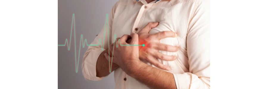
Shunt closures with device are a highly effective, minimally invasive treatment option for various congenital heart defects that involve abnormal blood flow between the heart chambers or vessels. These defects, such as Atrial Septal Defect (ASD), Ventricular Septal Defect (VSD), Patent Ductus Arteriosus (PDA), Ruptured Sinus of Valsalva (RSOV), and Aortopulmonary (AP) Window, can cause a range of symptoms and complications if left untreated. Dr. Akshat Jain, a renowned interventional cardiologist, specializes in the use of device closure techniques to treat these defects, offering patients a safer, faster recovery alternative to traditional open-heart surgery.

What Are Shunt Closures with Device?

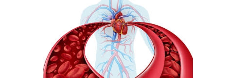
Shunt closures with device involve the percutaneous placement of a device to close abnormal openings (shunts) between the heart chambers or vessels. These devices are designed to seal off the defect, preventing blood from flowing abnormally between the left and right sides of the heart, or between the aorta and pulmonary artery, which is crucial for normal heart function.

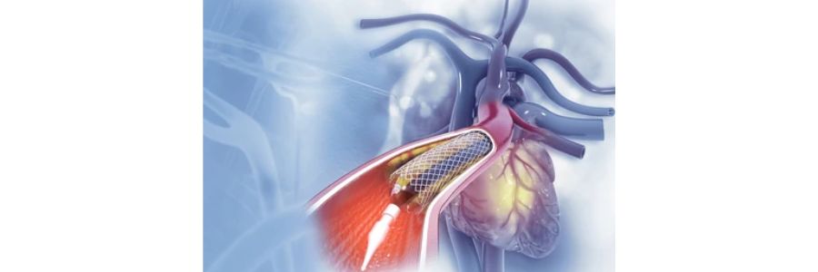
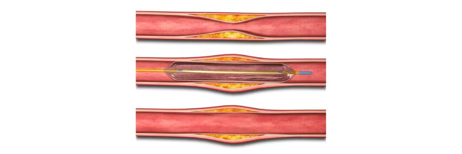
Shunt closure is typically performed using catheter-based techniques, where a device is inserted through a blood vessel (usually in the groin) and guided to the heart using advanced imaging technology like fluoroscopy and echocardiography. Once positioned correctly, the device expands to seal the defect, and over time, the body forms tissue around it, effectively closing the abnormal passage.

Types of Shunt Closures with Device

1. Atrial Septal Defect (ASD) Closure

Atrial Septal Defect (ASD) is a hole in the septum (wall) that separates the left and right atria. This defect allows oxygen-rich blood from the left atrium to flow into the right atrium, leading to inefficient blood circulation. Symptoms may include fatigue, palpitations, and shortness of breath.

Device Closure for ASD involves using a special umbrella-like device to close the hole. This procedure can often be done with minimal sedation and is typically performed in a catheterization lab. Device closure prevents complications like stroke or pulmonary hypertension that can arise from untreated ASD.

2. Ventricular Septal Defect (VSD) Closure

Ventricular Septal Defect (VSD) is a hole in the septum between the left and right ventricles. This defect causes oxygen-rich and oxygen-poor blood to mix, putting strain on the heart and potentially leading to heart failure or pulmonary hypertension if left untreated.

Device Closure for VSD uses a self-expanding occlusion device that is delivered via a catheter to the defect site. The device seals the VSD, preventing the abnormal blood flow and improving the heart's function.

3. Patent Ductus Arteriosus (PDA) Closure

Patent Ductus Arteriosus (PDA) is a persistent opening between the aorta and the pulmonary artery that should normally close after birth. If it remains open, it can lead to excessive blood flow to the lungs, potentially causing heart failure or pulmonary hypertension.

Device Closure for PDA involves placing a coil or occlusion device to close the abnormal duct. This procedure is typically performed in infants or children, but it can also be used for adults who have an undiagnosed or untreated PDA.

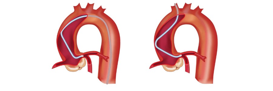
4. Ruptured Sinus of Valsalva (RSOV) Closure

Ruptured Sinus of Valsalva (RSOV) is a rare but serious heart condition where a tear or rupture occurs in one of the sinuses of Valsalva, which are the small pockets located behind the aortic valve. This rupture allows blood to flow from the aorta into the heart chambers, typically the right atrium or ventricle, leading to symptoms like chest pain, shortness of breath, fatigue, and in severe cases, heart failure.

This condition can be congenital or acquired, often resulting from an underlying structural heart disease or trauma. If left untreated, a ruptured sinus of Valsalva can cause significant complications, including cardiac tamponade or pulmonary hypertension. Treatment typically involves surgical repair or, in some cases, endovascular procedures to close the rupture. Timely diagnosis and intervention are crucial to prevent life-threatening complications.

5. Aortopulmonary (AP) Window Closure

Aortopulmonary (AP) Window is a rare congenital defect where an abnormal connection exists between the aorta and the pulmonary artery, allowing oxygen-rich and oxygen-poor blood to mix. This condition can result in heart failure or pulmonary hypertension.

Device Closure for AP Window involves the insertion of a specialized device to close the abnormal connection. By closing this window, the procedure improves circulation and reduces strain on the heart and lungs.

Why Choose Shunt Closure with Device?

  • Minimally Invasive: Shunt closure with device is a non-surgical procedure that requires only a small incision, typically in the groin, for catheter access, minimizing the need for large incisions or prolonged recovery.
  • Quick Recovery: Most patients can go home within a day or two and resume normal activities after a short recovery period, with minimal pain or discomfort.
  • Reduced Risk of Complications: Device closure reduces the risk of heart failure, stroke, or pulmonary hypertension, leading to better long-term health outcomes for patients.
  • Effective Long-Term Results: The device, once implanted, provides a permanent solution to the defect, with high success rates and minimal risk of recurrence.

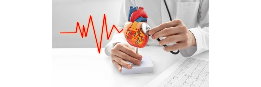
Why Choose Dr. Akshat Jain for Shunt Closure with Device?

  • Expert in Interventional Cardiology: Dr. Akshat Jain is a highly skilled interventional cardiologist with years of experience in performing complex catheter-based procedures, including shunt closures.
  • State-of-the-Art Technology: Dr. Jain uses the latest imaging and device technologies to perform precise and safe procedures, ensuring the best outcomes for patients.
  • Patient-Centered Care: Dr. Jain tailors each treatment plan to the specific needs of the patient, ensuring personalized care and optimal results.
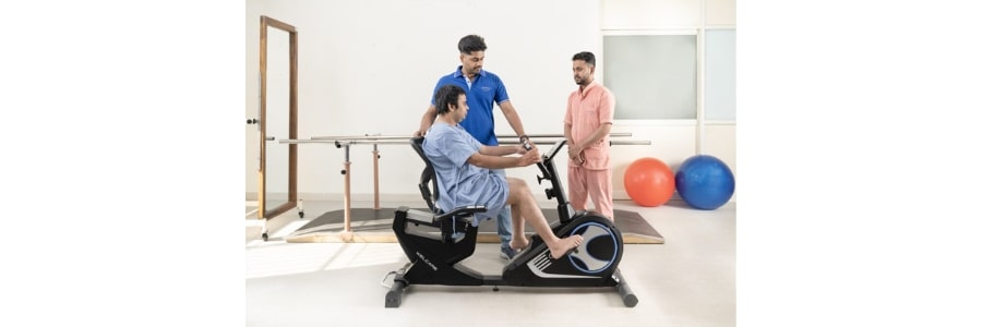
  • Comprehensive Follow-Up: Post-procedure care is crucial for ensuring that the device is functioning correctly. Dr. Jain offers thorough follow-up to monitor recovery and heart function.

Restore Your Heart Health with Shunt Closure

If you or a loved one has been diagnosed with a congenital heart defect, shunt closure with device may provide a safe, effective solution to restore normal heart function. Dr. Akshat Jain offers advanced interventional techniques to treat ASD, VSD, PDA, RSOV, and AP Window, helping you regain optimal heart health.

Contact Dr. Akshat Jain today to learn more about this life-changing procedure and take the first step toward a healthier heart.

Pulmonary Embolectomy: A Life-Saving Procedure for Pulmonary Embolism

Pulmonary embolectomy is a critical surgical procedure performed to treat pulmonary embolism (PE), a life-threatening condition where a blood clot (embolus) blocks one or more arteries in the lungs, disrupting blood flow and potentially leading to respiratory and cardiac failure. Dr. Akshat Jain, a skilled interventional cardiologist, specializes in advanced techniques, including pulmonary embolectomy, to manage this severe condition and restore blood flow to the lungs.

What is Pulmonary Embolism (PE)?

Pulmonary embolism occurs when a blood clot, often originating in the deep veins of the legs (deep vein thrombosis, or DVT), breaks loose and travels through the bloodstream to the lungs. The clot can lodge in one of the pulmonary arteries, blocking the flow of oxygenated blood and causing potentially severe complications, such as:

  • Hypoxia (low oxygen levels in the blood)
  • Right-sided heart failure
  • Shock
  • Cardiac arrest

PE is a medical emergency that requires immediate treatment to prevent life-threatening complications. Depending on the severity of the blockage, pulmonary embolectomy may be considered as a life-saving intervention.

When is Pulmonary Embolectomy Performed?

Pulmonary embolectomy is typically recommended in the following situations:

  • Massive Pulmonary Embolism: When a large clot obstructs the pulmonary artery, leading to a significant drop in blood pressure and causing shock or respiratory failure.
  • Failure of Other Treatments: If other methods, such as thrombolysis (clot-busting drugs) or catheter-based therapies, do not effectively resolve the blockage.
  • High Risk of Death: In patients with severe PE who are at immediate risk of death and are not candidates for thrombolysis or catheter interventions.

The decision to perform pulmonary embolectomy is based on the patient’s condition, severity of the embolism, and overall health. Dr. Akshat Jain carefully evaluates each case to determine the most appropriate treatment option.

How is Pulmonary Embolectomy Performed?

1. Surgical Pulmonary Embolectomy:

  • This involves making an incision in the chest and directly accessing the pulmonary arteries to remove the clot.
  • It is performed in more severe cases where other methods have not been effective.

2. Catheter-Directed Pulmonary Embolectomy:

  • This minimally invasive technique involves the insertion of a catheter through a vein, usually in the groin or neck, to reach the pulmonary arteries.
  • The catheter is used to remove or break up the clot using mechanical devices or suction.

The procedure aims to quickly restore normal blood flow to the lungs, reducing the risk of permanent damage to the heart and lungs.

Post-Procedure Care and Recovery

After undergoing pulmonary embolectomy, patients are closely monitored in an intensive care unit (ICU) setting for several days. The recovery process includes:

  • Monitoring vital signs: Such as blood pressure, heart rate, and oxygen levels.
  • Respiratory support: If necessary, to ensure proper oxygenation.
  • Pain management: To ease discomfort from the procedure.
  • Anticoagulant therapy: To prevent the formation of new clots and reduce the risk of further embolic events.
  • Gradual mobilization: To prevent complications from prolonged bed rest.

The recovery time can vary based on the severity of the pulmonary embolism and the patient's overall health. Most patients can return to normal activities within a few weeks, although regular follow-up care is necessary to monitor for any potential complications.

Why Choose Dr. Akshat Jain for Pulmonary Embolectomy?

  • Expertise in Critical Care: Dr. Akshat Jain is highly skilled in managing complex cardiovascular conditions, including pulmonary embolism. He uses advanced techniques to ensure the best outcomes for his patients.
  • Personalized Treatment: Each pulmonary embolism case is unique, and Dr. Jain customizes the treatment approach based on the individual’s condition and medical history.
  • Comprehensive Care: From diagnosis to post-procedure care, Dr. Jain provides comprehensive management, including preventive strategies to reduce the risk of recurrent pulmonary embolism.

Preventing Pulmonary Embolism

While pulmonary embolectomy is a life-saving procedure, preventing pulmonary embolism is crucial. Dr. Akshat Jain emphasizes the importance of:

  • Early detection and treatment of deep vein thrombosis (DVT) with anticoagulant medications.
  • Mobilization after surgery or long periods of immobility to promote healthy circulation.
  • Compression stockings for patients at risk of blood clots.
  • Regular monitoring for individuals with known risk factors such as cancer, obesity, or recent surgery.

By managing risk factors and taking proactive measures, the incidence of PE can be significantly reduced.

Conclusion

Pulmonary embolectomy is a crucial procedure for managing severe cases of pulmonary embolism, restoring blood flow to the lungs, and saving lives. Dr. Akshat Jain's expertise in interventional cardiology and cardiovascular procedures ensures that patients receive the highest level of care, tailored to their specific needs. If you or a loved one is at risk for pulmonary embolism or experiencing symptoms of PE, seeking early medical attention is essential.

Schedule a consultation with Dr. Akshat Jain today to learn more about pulmonary embolism prevention and treatment options.

Coarctoplasty: A Minimally Invasive Treatment for Coarctation of the Aorta

Coarctation of the aorta (CoA) is a congenital condition where a narrowing of the aorta restricts blood flow, leading to elevated blood pressure and increased workload on the heart. If left untreated, CoA can result in serious complications, including heart failure, stroke, and kidney damage. Coarctoplasty, a minimally invasive procedure performed by Dr. Akshat Jain, is a highly effective treatment for this condition, offering patients a safe and efficient way to restore normal aortic function and improve heart health.

What is Coarctoplasty?

Coarctoplasty is a non-surgical procedure used to treat aortic coarctation by widening the narrowed section of the aorta. This procedure can be performed using a balloon catheter or a stent, depending on the severity and location of the narrowing. By restoring normal blood flow through the aorta, coarctoplasty helps reduce the strain on the heart and lowers blood pressure, significantly improving the patient’s quality of life and long-term health outcomes.

How is Coarctoplasty Performed?

1. Preparation:

  • The procedure is typically performed under local anesthesia and sedation.
  • A catheter is inserted into a blood vessel, usually in the groin, and guided to the site of the coarctation in the aorta.

Balloon Angioplasty (Balloon Coarctoplasty):

  • In the most common form of coarctoplasty, a balloon catheter is inserted into the narrowed portion of the aorta and inflated to widen the constricted area.
  • This helps restore normal blood flow by stretching the narrowed segment of the artery.

Stent Placement:

  • In some cases, a stent may be placed to keep the aorta open after balloon angioplasty, especially if the coarctation is severe or at risk of recurring.

Post-Procedure Monitoring:

  • After the procedure, patients are monitored to ensure that the procedure was successful and to check for any complications, such as bleeding or re-narrowing of the aorta.

Why is Coarctoplasty Important?

Coarctoplasty offers several benefits that are crucial for patients with aortic coarctation:

  • Improved Blood Flow: Coarctoplasty effectively relieves the narrowing of the aorta, allowing for normal blood flow and reducing the strain on the heart.
  • Lower Blood Pressure: By eliminating the obstruction, the procedure helps lower high blood pressure, which is common in patients with coarctation of the aorta, reducing the risk of complications like stroke, heart failure, and kidney damage.
  • Minimally Invasive: Coarctoplasty is performed with a catheter, meaning no open surgery is required. This results in a quicker recovery and fewer risks compared to traditional surgical options.
  • Long-Term Benefits: For many patients, coarctoplasty offers a permanent solution to the condition, improving overall heart function and reducing the need for further interventions.

When is Coarctoplasty Recommended?

Coarctoplasty is typically recommended for patients diagnosed with aortic coarctation who experience:

  • High blood pressure that is difficult to control with medication
  • Chest pain, shortness of breath, or symptoms of heart failure
  • Poor exercise tolerance due to restricted blood flow
  • Signs of organ damage, such as kidney problems

It is also recommended for individuals with coarctation who have not responded to other treatments or medications and those who are not candidates for open surgery.

Why Choose Dr. Akshat Jain for Coarctoplasty?

  • Expert in Interventional Cardiology: Dr. Akshat Jain has extensive experience in performing complex, minimally invasive procedures like coarctoplasty, ensuring the highest level of precision and care.
  • State-of-the-Art Technology: Using advanced imaging techniques, Dr. Jain ensures the procedure is carried out safely and effectively, targeting the narrowed portion of the aorta with minimal risk of complications.
  • Comprehensive Care: Dr. Jain provides personalized treatment plans, tailored to each patient’s specific needs and health status, with ongoing monitoring and follow-up care.
  • Minimized Recovery Time: With coarctoplasty, patients typically experience a faster recovery compared to open surgery, allowing them to return to normal activities with reduced risk of complications.

Take Control of Your Heart Health with Coarctoplasty

If you or a loved one has been diagnosed with coarctation of the aorta, coarctoplasty may be the right solution. This minimally invasive procedure offers a safe and effective way to restore normal blood flow, reduce high blood pressure, and improve heart function.

Contact Dr. Akshat Jain today to schedule a consultation and learn more about how coarctoplasty can improve your heart health and overall well-being.

Balloon Mitral Valvuloplasty (BMV): A Minimally Invasive Solution for Mitral Stenosis

Balloon Mitral Valvuloplasty (BMV) is an advanced, minimally invasive procedure used to treat mitral stenosis, a condition where the mitral valve becomes narrowed, restricting blood flow from the left atrium to the left ventricle. By restoring proper valve function, BMV improves blood flow, relieves symptoms, and enhances the patient’s quality of life.

What is Mitral Stenosis?

Mitral stenosis occurs when the mitral valve leaflets become thickened or fused, limiting their ability to open fully. Common symptoms include:

  • Shortness of breath
  • Fatigue
  • Swelling in the legs or abdomen
  • Palpitations or irregular heartbeats

If left untreated, mitral stenosis can lead to complications such as heart failure, pulmonary hypertension, or stroke.

What is Balloon Mitral Valvuloplasty?

BMV, also known as Percutaneous Mitral Commissurotomy, is a catheter-based procedure designed to widen the narrowed mitral valve without the need for open-heart surgery. It involves the use of a specialized balloon catheter to separate the fused valve leaflets, restoring normal blood flow.

Who is a Candidate for BMV?

BMV is ideal for patients with:

  • Rheumatic Mitral Stenosis: The most common cause of mitral valve narrowing.
  • Symptomatic Mitral Stenosis: Experiencing shortness of breath, fatigue, or other signs of reduced blood flow.
  • Favorable Valve Anatomy: Based on echocardiographic evaluation.

Patients with heavily calcified valves or significant mitral regurgitation may not be suitable for BMV and might require alternative treatments.

The BMV Procedure

1. Preparation:

  • The procedure is performed under local anesthesia and mild sedation in a cath lab.
  • Imaging tools like fluoroscopy and echocardiography guide the process.

2. Catheter Insertion:

  • A catheter is introduced through a vein in the groin and guided to the heart.
  • A small puncture is made in the atrial septum (a thin wall between the heart's upper chambers) to access the mitral valve.

3. Balloon Inflation:

  • A specialized balloon catheter is positioned within the narrowed mitral valve.
  • The balloon is inflated, gently separating the fused valve leaflets to increase the valve’s opening size.

4. Completion:

  • The balloon is deflated and removed, and the catheter is withdrawn.

The entire procedure typically takes 1-2 hours, and patients often experience immediate symptom relief.

Benefits of Balloon Mitral Valvuloplasty

  • Minimally Invasive: No need for open-heart surgery or prolonged hospital stays.
  • Quick Recovery: Most patients return home within 24-48 hours.
  • Immediate Symptom Relief: Enhanced blood flow reduces breathlessness and fatigue.
  • Improved Quality of Life: Enables patients to resume daily activities with ease.
  • Safe and Effective: High success rates in eligible patients.

Recovery and Aftercare

  • Hospital Stay: Typically discharged within 1-2 days.
  • Activity: Resume light activities within a few days; avoid strenuous tasks for a couple of weeks.
  • Medications: Continue prescribed medications to manage symptoms or prevent blood clots.
  • Follow-Up: Regular check-ups and echocardiograms to monitor valve function.

Why Choose Us for Balloon Mitral Valvuloplasty?

  • Expert Cardiologist: Our skilled interventional cardiologist specializes in advanced valve procedures.
  • State-of-the-Art Facilities: Equipped with cutting-edge technology for precise and safe interventions.
  • Comprehensive Care: From evaluation to post-procedure follow-up, we ensure personalized treatment tailored to your needs.

Is BMV the Right Option for You?

If you or a loved one has been diagnosed with mitral stenosis, Balloon Mitral Valvuloplasty could be the minimally invasive solution you’ve been searching for. Contact our expert Dr. Akshat Jain today to schedule a consultation and learn more about this life-changing procedure.

Balloon Pulmonic Valvuloplasty (BPV): A Minimally Invasive Treatment for Pulmonary Valve Stenosis

Balloon Pulmonic Valvuloplasty (BPV) is a specialized, minimally invasive procedure used to treat pulmonary valve stenosis, a condition where the pulmonary valve becomes narrowed, obstructing blood flow from the right ventricle to the lungs. BPV effectively widens the valve, improving blood flow and alleviating symptoms without the need for open-heart surgery.

Understanding Pulmonary Valve Stenosis

Pulmonary valve stenosis occurs when the pulmonary valve’s leaflets thicken, stiffen, or fuse together, limiting their ability to open fully. This increases the workload on the right ventricle, potentially leading to symptoms such as:

  • Fatigue
  • Shortness of breath
  • Chest pain
  • Fainting (syncope)
  • Heart murmur

If untreated, severe pulmonary valve stenosis can lead to complications such as right heart failure or arrhythmias.

What is Balloon Pulmonic Valvuloplasty?

BPV is a catheter-based procedure designed to improve blood flow by widening the narrowed pulmonary valve. A specialized balloon catheter is used to stretch and separate the valve leaflets, restoring proper valve function.

Who is a Candidate for BPV?

BPV is recommended for patients with:

  • Moderate to Severe Pulmonary Valve Stenosis: With symptoms or evidence of heart strain.
  • Congenital Heart Defects: Pulmonary valve abnormalities present since birth.
  • Right Ventricular Hypertrophy: Thickening of the right ventricle due to increased workload.

Patients with significant pulmonary regurgitation or other complex congenital heart conditions may require alternative treatments.

The BPV Procedure

1. Preparation:

  • BPV is performed under local anesthesia and mild sedation or general anesthesia in a cardiac catheterization lab.
  • Imaging techniques like echocardiography and fluoroscopy guide the procedure.

2. Catheter Insertion:

  • A catheter is introduced through a vein in the groin or neck and guided to the pulmonary valve.

3. Balloon Inflation:

  • A deflated balloon catheter is positioned within the narrowed valve.
  • The balloon is inflated to stretch the valve and separate the fused leaflets.

4. Completion:

  • The balloon is deflated and removed, and the catheter is withdrawn.
  • The procedure typically lasts 1-2 hours, and most patients experience immediate improvement in symptoms.

Benefits of Balloon Pulmonic Valvuloplasty

  • Minimally Invasive: No need for open-heart surgery.
  • Short Recovery Time: Most patients return home within a day or two.
  • Immediate Results: Significant improvement in valve function and symptom relief.
  • Low Risk: Safe and effective with minimal complications in eligible patients.
  • Durable Outcomes: Long-lasting improvement in blood flow for most patients.

Recovery and Aftercare

  • Hospital Stay: Patients are typically monitored overnight and discharged the next day.
  • Activity: Light activities can be resumed within a few days; strenuous tasks should be avoided for 1-2 weeks.
  • Follow-Up Care: Regular follow-ups with echocardiograms to monitor valve function.

Why Choose Us for BPV?

  • Expert Cardiologist: Our experienced interventional cardiologist specializes in valve procedures, ensuring precision and safety.
  • Advanced Facilities: Equipped with state-of-the-art technology for accurate diagnosis and treatment.
  • Comprehensive Care: From initial evaluation to post-procedure follow-ups, we provide personalized, patient-focused care.

Is BPV the Right Choice for You? If you or a loved one has been diagnosed with pulmonary valve stenosis, Balloon Pulmonic Valvuloplasty offers a safe, effective, and minimally invasive treatment option to restore valve function and improve quality of life. Contact our expert Dr. Akshat Jain today to learn more about BPV and schedule your consultation.

Alcohol Septal Ablation for Hypertrophic Cardiomyopathy: A Minimally Invasive Treatment Option

Alcohol Septal Ablation (ASA) is a groundbreaking, minimally invasive procedure used to treat Hypertrophic Cardiomyopathy (HCM), a condition characterized by abnormal thickening of the heart muscle, particularly the septum (the wall separating the left and right sides of the heart). This thickening can obstruct blood flow and lead to symptoms like shortness of breath, chest pain, fainting, and, in severe cases, sudden cardiac death. Dr. Akshat Jain, a leading interventional cardiologist, specializes in performing Alcohol Septal Ablation to improve heart function and quality of life for patients with HCM who are not candidates for surgery.

What is Alcohol Septal Ablation?

Alcohol Septal Ablation is a non-surgical procedure that aims to reduce the thickness of the heart's septal wall by injecting alcohol into the coronary artery that supplies the thickened heart tissue. The alcohol causes a controlled, targeted ischemic injury (a lack of blood supply) to the thickened septal muscle, leading to its shrinkage and improved blood flow. This results in the reduction of the left ventricular outflow tract obstruction (LVOTO), which is the primary cause of symptoms in HCM patients.

Key Benefits of Alcohol Septal Ablation:

  • Minimally Invasive: Unlike traditional surgery, ASA is performed through a catheter inserted into the coronary arteries, requiring only a small incision in the groin or wrist.
  • Effective Symptom Relief: By reducing the hypertrophic tissue, ASA significantly improves symptoms like shortness of breath, chest pain, and fatigue, enhancing quality of life.
  • No Need for Open Surgery: Alcohol Septal Ablation offers an effective treatment option for patients who are not suitable candidates for traditional surgical procedures, such as myectomy.
  • Quick Recovery: The procedure is relatively quick, and most patients can return home the same day or the next, with a fast recovery time compared to open-heart surgery.

How is Alcohol Septal Ablation Performed?

1. Preparation:

  • The procedure is typically performed under local anesthesia and sedation.
  • A catheter is inserted into a blood vessel, usually in the groin or wrist, and guided to the coronary arteries using X-ray fluoroscopy.

2. Identification of the Target Area:

  • Using imaging techniques such as echocardiography and coronary angiography, the exact location of the thickened heart tissue and the coronary artery supplying it is identified.

3. Injection of Alcohol:

  • A small amount of alcohol (usually ethanol) is injected into the targeted coronary artery.
  • The alcohol causes a controlled injury to the heart muscle, reducing its thickness and relieving obstruction.

4. Monitoring and Follow-Up:

  • After the procedure, patients are monitored for a short period to ensure no complications arise.
  • Follow-up visits are scheduled to assess improvement in symptoms and heart function.

When is Alcohol Septal Ablation Recommended?

Alcohol Septal Ablation is typically considered for patients with Hypertrophic Cardiomyopathy who meet the following criteria:

  • Severe Symptoms: Patients experiencing significant symptoms like shortness of breath, chest pain, or fainting that do not improve with medications.
  • Obstructed Left Ventricular Outflow Tract (LVOT): ASA is particularly effective in treating obstructive hypertrophic cardiomyopathy (HCM), where the thickened septal tissue obstructs the flow of blood from the left ventricle.
  • Not a Surgical Candidate: For patients who are not candidates for surgical treatment, such as those with high surgical risk or multiple comorbidities, ASA provides a less invasive alternative.

Why Choose Dr. Akshat Jain for Alcohol Septal Ablation?

  • Expert in Interventional Cardiovascular Procedures: Dr. Akshat Jain has extensive experience in performing Alcohol Septal Ablation, ensuring precision and safety throughout the procedure.
  • Advanced Imaging Technology: The procedure is guided by the latest imaging techniques, including echocardiography and angiography, to ensure accurate placement of the catheter and alcohol injection.
  • Personalized Care: Dr. Jain takes a patient-centered approach, tailoring each treatment plan to the unique needs and medical history of the patient.
  • Comprehensive Follow-Up: After the procedure, Dr. Jain ensures thorough monitoring and follow-up care, assessing the patient's recovery and long-term success of the treatment.

Take the First Step Towards Better Heart Health. If you or a loved one is living with Hypertrophic Cardiomyopathy and struggling with symptoms that are affecting daily life, Alcohol Septal Ablation may provide an effective solution. With Dr. Akshat Jain, a leader in the field of interventional cardiology, you can receive top-tier care using the latest, minimally invasive treatments for HCM. Contact Dr. Akshat Jain, to learn more about Alcohol Septal Ablation and how it can help you regain control over your heart health.

Heart Failure Therapy: Comprehensive Management for Better Heart Health

Heart failure is a serious condition in which the heart is unable to pump blood efficiently to meet the body’s needs. It can occur due to various causes, including coronary artery disease, hypertension, and valve disorders. Heart failure can significantly impact a person’s quality of life, but with early diagnosis and appropriate treatment, patients can lead a better, healthier life. Dr. Akshat Jain, a leading interventional cardiologist, specializes in heart failure therapy, offering personalized treatment plans to optimize heart function and improve overall well-being.

What is Heart Failure?

Heart failure (HF) occurs when the heart is too weak or stiff to pump blood effectively, leading to fluid buildup in the lungs, abdomen, and other parts of the body. Common symptoms include:

  • Shortness of breath, especially during exertion
  • Fatigue and reduced exercise tolerance
  • Swelling in the legs, ankles, or abdomen
  • Persistent cough or wheezing
  • Rapid or irregular heartbeat

Heart failure can be categorized into left-sided heart failure, right-sided heart failure, or congestive heart failure, depending on which part of the heart is affected. It can also be classified by the heart's ability to pump blood, such as heart failure with reduced ejection fraction (HFrEF) or heart failure with preserved ejection fraction (HFpEF).

Heart Failure Therapy: Multidisciplinary Approach

The management of heart failure involves a combination of medical, interventional, and lifestyle therapies tailored to the individual patient. Dr. Akshat Jain focuses on offering advanced heart failure therapies that aim to improve heart function, alleviate symptoms, and prevent disease progression.

1. Medical Management of Heart Failure

  • Medications: The cornerstone of heart failure therapy includes several classes of medications designed to help the heart pump more effectively, reduce fluid buildup, and lower blood pressure:
    • ACE inhibitors or ARBs to relax blood vessels and reduce strain on the heart
    • Beta-blockers to control heart rate and improve heart pumping efficiency
    • Diuretics to help the body get rid of excess fluid
    • Aldosterone antagonists to prevent harmful effects of high aldosterone levels
    • ARNI (Angiotensin Receptor-Neprilysin Inhibitors) for advanced heart failure management
    • SGLT2 inhibitors to reduce hospitalization risk in heart failure patients
  • Lifestyle Modifications: In addition to medication, heart failure patients are advised to make lifestyle changes, such as:
    • Maintaining a low-sodium diet to reduce fluid retention
    • Regular exercise tailored to the patient’s condition to strengthen the heart
    • Weight management to reduce strain on the heart
    • Limiting alcohol and avoiding smoking to improve overall cardiovascular health

2. Interventional Heart Failure Therapies

While medical management is critical, interventional procedures can also play a key role in treating heart failure, especially in patients with advanced disease or complications like valve issues or coronary artery disease.

a. Percutaneous Coronary Interventions (PCI):

For heart failure patients with coronary artery disease (CAD), angioplasty or stenting may be required to restore blood flow to the heart muscle. Dr. Akshat Jain specializes in advanced PCI techniques to improve coronary circulation, which can reduce the burden on the heart and alleviate symptoms of heart failure.

b. Cardiac Resynchronization Therapy (CRT):

In heart failure patients with dyssynchronous heart contraction, Cardiac Resynchronization Therapy (CRT), also known as biventricular pacing, can improve heart function by synchronizing the heart's contractions. This therapy involves implanting a specialized pacemaker that coordinates the heart’s rhythm, reducing symptoms and improving exercise capacity.

c. Implantable Cardioverter Defibrillator (ICD):

For patients with heart failure at risk of life-threatening arrhythmias, an ICD can be implanted to monitor the heart’s rhythm and deliver shocks if necessary to restore normal rhythm. This device can significantly reduce the risk of sudden cardiac death in heart failure patients.

d. Left Ventricular Assist Device (LVAD):

In cases of severe heart failure, especially when the heart can no longer pump effectively, a Left Ventricular Assist Device (LVAD) may be implanted to help the heart pump blood. LVADs can be a bridge to heart transplantation or, in some cases, a long-term solution for patients who are not candidates for a transplant.

3. Advanced Therapies for Severe Heart Failure

In cases of end-stage heart failure, when conventional treatments are no longer effective, more advanced therapies may be considered:

a. Heart Transplantation:

For some patients, a heart transplant may be the only viable option when all other treatments have failed. This involves replacing the diseased heart with a healthy donor heart.

b. Alcohol Septal Ablation for Hypertrophic Cardiomyopathy (HCM):

For patients with hypertrophic cardiomyopathy (HCM), a condition that can contribute to heart failure, Alcohol Septal Ablation is a minimally invasive procedure that targets and reduces the thickened heart muscle, improving blood flow and relieving symptoms.

Why Choose Dr. Akshat Jain for Heart Failure Therapy?

  • Expert in Advanced Interventions: Dr. Akshat Jain is an experienced interventional cardiologist specializing in the management of complex heart failure cases using both medical and interventional therapies.
  • Personalized Treatment Plans: Dr. Jain takes a holistic approach to heart failure management, customizing treatment plans based on each patient’s unique needs, health status, and goals.
  • Cutting-Edge Technologies: Using the latest diagnostic tools, imaging techniques, and therapies, Dr. Jain ensures optimal results for heart failure patients, improving both quality of life and long-term outcomes.
  • Comprehensive Care: From diagnosis through long-term management, Dr. Jain offers continuous support and follow-up care to ensure the best possible outcomes for heart failure patients.

Take Charge of Your Heart Health

Heart failure is a serious condition, but with the right treatment approach, it is possible to manage symptoms, improve heart function, and lead a fulfilling life. Dr. Akshat Jain offers comprehensive heart failure therapy, combining advanced interventions, medications, and lifestyle changes to improve heart health and enhance your quality of life.

Contact Dr. Akshat Jain today to learn more about heart failure management and explore the best treatment options available for you.

Pulmonary Hypertension Management: Comprehensive Care for a Complex Condition

Pulmonary hypertension (PH) is a serious condition where the blood pressure in the arteries of the lungs becomes abnormally high, putting strain on the heart and leading to progressive heart failure. Managing pulmonary hypertension requires a multi-disciplinary approach, combining medical treatment, lifestyle modifications, and advanced interventional techniques. Dr. Akshat Jain, an expert interventional cardiologist, offers comprehensive care for pulmonary hypertension, helping patients lead healthier and more fulfilling lives.

What is Pulmonary Hypertension?

Pulmonary hypertension occurs when there is an increase in the pressure within the pulmonary arteries, the blood vessels that carry blood from the heart to the lungs. This elevated pressure makes it more difficult for the heart, particularly the right ventricle, to pump blood effectively to the lungs. As a result, the heart works harder, leading to symptoms such as:

  • Shortness of breath
  • Fatigue
  • Chest pain
  • Dizziness or fainting
  • Swelling in the ankles, legs, or abdomen

Pulmonary hypertension can be caused by a variety of conditions, including:

  • Chronic lung diseases (e.g., COPD, interstitial lung disease)
  • Heart conditions (e.g., left heart disease, congenital heart defects)
  • Chronic blood clots in the lungs (pulmonary embolism)
  • Connective tissue diseases (e.g., scleroderma, lupus)
  • Idiopathic pulmonary arterial hypertension (in cases with no known cause)

Phases of Pulmonary Hypertension Management

Managing pulmonary hypertension requires an individualized approach that addresses the root cause, relieves symptoms, and improves overall cardiovascular health. Dr. Akshat Jain follows a structured approach that includes the following phases:

1. Early Diagnosis and Evaluation

The first step in managing pulmonary hypertension is early detection. Early symptoms can be subtle, making it essential to identify and diagnose the condition before complications arise. Diagnostic tools used in assessing PH include:

  • Echocardiogram: A non-invasive ultrasound that evaluates the heart’s function and can suggest increased pulmonary pressure.
  • Right heart catheterization: The gold standard for confirming the diagnosis by directly measuring the pressure in the pulmonary arteries.
  • Pulmonary function tests: To assess lung capacity and function.
  • CT pulmonary angiography: Used to assess for chronic blood clots or lung disease.
  • Blood tests: To identify underlying causes such as autoimmune diseases or infections.

Once diagnosed, Dr. Jain creates a personalized treatment plan based on the underlying cause of PH, the severity of symptoms, and the overall health of the patient.

2. Medical Management

The goal of medical treatment is to manage symptoms, slow disease progression, and improve quality of life. Dr. Jain employs a range of medications, depending on the type of pulmonary hypertension:

  • Vasodilators: Medications such as prostacyclin analogs, endothelin receptor antagonists, and phosphodiesterase inhibitors can relax and dilate the pulmonary arteries, lowering blood pressure in the lungs.
  • Anticoagulants: Blood thinners may be prescribed to prevent blood clots from forming in patients with chronic thromboembolic pulmonary hypertension (CTEPH).
  • Diuretics: These help reduce fluid retention and relieve swelling caused by right heart failure.
  • Oxygen therapy: For patients with low blood oxygen levels, supplemental oxygen helps improve breathing and oxygenation.
  • Heart failure management: For those with right-sided heart failure, medications like ACE inhibitors, beta-blockers, or diuretics may be used.

Medications are often tailored to the specific subtype of pulmonary hypertension and adjusted based on patient response.

3. Interventional Treatments

In certain cases, when medical management alone is insufficient, interventional treatments may be required to improve blood flow and reduce pulmonary pressures. Dr. Akshat Jain is skilled in advanced interventions for PH management, including:

  • Balloon Pulmonary Angioplasty (BPA): For patients with chronic thromboembolic pulmonary hypertension (CTEPH), this minimally invasive procedure involves inflating a balloon inside the blocked pulmonary arteries to open them and improve blood flow.
  • Surgical Pulmonary Thromboendarterectomy (PTE): In more severe cases of CTEPH, surgery to remove the blood clots from the pulmonary arteries may be necessary.
  • Right heart catheterization and testing: Dr. Jain may use catheter-based techniques for diagnostic or therapeutic purposes to measure pressures directly and determine the best treatment strategy.

4. Lifestyle Modifications and Supportive Care

Living with pulmonary hypertension requires careful attention to overall lifestyle. Dr. Jain emphasizes the importance of lifestyle changes that help manage PH, including:

  • Exercise programs: Supervised exercise can help improve overall cardiovascular fitness, lung function, and quality of life.
  • Dietary adjustments: A heart-healthy diet, including low-sodium meals, can help manage symptoms of right heart failure and prevent fluid retention.
  • Smoking cessation: If the patient is a smoker, quitting is essential to prevent further damage to the lungs and heart.
  • Psychological support: Living with a chronic illness can lead to emotional challenges. Dr. Jain provides counseling or refers patients to support groups to help manage the emotional burden of pulmonary hypertension.

5. Long-Term Monitoring and Follow-Up

Pulmonary hypertension is a chronic condition that requires long-term monitoring to assess the effectiveness of treatment and prevent complications. Regular follow-up visits with Dr. Akshat Jain ensure the patient’s progress is closely monitored, with adjustments made to the treatment plan as needed. Monitoring includes:

  • Repeat echocardiograms to assess heart function.
  • Pulmonary function tests to track lung health.
  • Blood tests to evaluate kidney function and potential side effects of medications.
  • Adjusting medications based on the patient’s condition and symptoms.

Why Choose Dr. Akshat Jain for Pulmonary Hypertension Management?

  • Expert Diagnosis and Tailored Treatment Plans: Dr. Akshat Jain utilizes state-of-the-art diagnostic techniques and creates individualized treatment plans that address the underlying cause and severity of pulmonary hypertension.
  • Comprehensive Care: From medication management to advanced interventional therapies, Dr. Jain offers a holistic approach to managing pulmonary hypertension, ensuring the best possible outcomes for his patients.
  • Innovative Interventions: As an experienced interventional cardiologist, Dr. Jain is skilled in advanced treatments such as balloon pulmonary angioplasty and right heart catheterization, offering patients cutting-edge therapies that can significantly improve their condition.

Conclusion

Pulmonary hypertension is a complex and potentially life-threatening condition, but with early diagnosis, comprehensive treatment, and ongoing management, individuals can lead more fulfilling and active lives. If you or a loved one is experiencing symptoms of pulmonary hypertension or has been diagnosed with the condition, Dr. Akshat Jain is dedicated to providing the highest level of care, from medical management to advanced interventional options.

Contact Dr. Akshat Jain’s clinic today to schedule a consultation and take the first step toward better heart and lung health.

Cardiac Rehabilitation: A Vital Step Toward Heart Health Recovery

Cardiac rehabilitation is a medically supervised program designed to help individuals recover from heart-related conditions, improve their cardiovascular health, and reduce the risk of future heart problems. Whether you're recovering from a heart attack, heart surgery, or managing chronic heart conditions, Dr. Akshat Jain offers comprehensive cardiac rehabilitation services tailored to each patient’s needs, helping them regain their strength and enjoy a healthier, active life.

What is Cardiac Rehabilitation?

Cardiac rehabilitation is a multi-phase program that combines exercise training, education, and counseling to help individuals with heart disease recover and improve their overall health. It is an essential component of heart disease management, helping patients reduce the risk of heart attacks, strokes, and other complications, while improving their quality of life.

Cardiac rehabilitation typically involves:

  • Exercise therapy: to strengthen the heart and improve physical fitness
  • Nutritional counseling: to support heart health and manage weight
  • Behavioral counseling: to address lifestyle changes such as smoking cessation, stress management, and emotional well-being
  • Education: to help patients understand their condition and how to manage it effectively

Benefits of Cardiac Rehabilitation

Cardiac rehabilitation offers numerous benefits that can help you recover more effectively and prevent future heart problems:

  • Improved heart function: Regular exercise and a healthy lifestyle can strengthen the heart and improve its efficiency.
  • Reduced risk of future heart events: By managing risk factors such as high blood pressure, high cholesterol, and diabetes, cardiac rehab reduces the chances of future heart attacks or strokes.
  • Improved quality of life: Patients who participate in cardiac rehabilitation often report feeling more energetic, less fatigued, and more confident about their heart health.
  • Weight management: Exercise and nutrition guidance can help patients achieve and maintain a healthy weight, reducing strain on the heart.
  • Psychological benefits: Cardiac rehabilitation also addresses the emotional aspects of heart disease, offering counseling and support to manage stress, anxiety, and depression.

Phases of Cardiac Rehabilitation

Cardiac rehabilitation typically consists of three phases:

Phase I: In-Hospital Rehabilitation

The first phase of cardiac rehabilitation starts during the hospital stay, immediately following a heart event or surgery (e.g., heart attack, coronary artery bypass surgery, or stent placement). During this phase, patients are closely monitored, and light activities, such as walking and breathing exercises, are introduced to begin the recovery process.

Phase II: Outpatient Rehabilitation

Phase II begins once the patient is discharged from the hospital and continues for several weeks to months. During this phase, patients engage in:

  • Supervised exercise programs tailored to their individual health status and recovery progress
  • Education sessions on heart-healthy diets, medications, and lifestyle changes
  • Stress management and counseling to promote emotional well-being

The goal of this phase is to help patients regain strength, build stamina, and learn long-term habits for heart health.

Phase III: Maintenance Phase

Phase III is the long-term maintenance phase, designed to help patients continue their progress independently. While it may involve less direct medical supervision, it includes regular exercise, weight management, and ongoing counseling to keep the heart healthy and prevent future issues.

How Cardiac Rehabilitation Works

Dr. Akshat Jain's approach to cardiac rehabilitation is comprehensive and personalized. The program includes:

  • Initial Evaluation: Dr. Jain will assess your heart health, current fitness level, medical history, and any other risk factors to design a personalized rehab plan.
  • Supervised Exercise: A customized exercise program will be designed to improve cardiovascular fitness, strength, and flexibility. The exercises will be adapted based on your condition and abilities, progressing as your fitness improves.
  • Nutritional Counseling: Dr. Jain's team will guide you on heart-healthy eating habits, helping you make better dietary choices that support cardiovascular health and weight management.
  • Behavioral Counseling: Emotional support, including strategies to reduce stress and anxiety, is offered to help you cope with the emotional challenges of heart disease recovery.
  • Continuous Monitoring: Your progress will be monitored regularly to ensure the rehabilitation program is effective and to adjust it as needed.

Who Should Consider Cardiac Rehabilitation?

Cardiac rehabilitation is beneficial for individuals who:

  • Have recently experienced a heart attack or heart surgery (bypass surgery, stent placement)
  • Are managing chronic heart conditions such as coronary artery disease or heart failure
  • Have undergone valve surgery or a heart transplant
  • Are recovering from procedures like angioplasty or percutaneous coronary intervention (PCI)
  • Have risk factors for heart disease, such as high blood pressure, diabetes, or high cholesterol

Even if you are not currently experiencing symptoms, preventive cardiac rehabilitation can be a valuable program for those looking to improve heart health and reduce risk factors for future heart problems.

Why Choose Dr. Akshat Jain for Cardiac Rehabilitation?

  • Expert Cardiologist: Dr. Akshat Jain brings years of experience in interventional cardiology and heart disease management. His expertise allows him to create tailored cardiac rehabilitation plans to meet the unique needs of each patient.
  • Personalized Care: Every patient receives an individualized rehabilitation plan, taking into account their medical history, heart condition, and recovery goals.
  • Comprehensive Support: Dr. Jain’s approach includes not only medical management but also psychological, dietary, and fitness support to ensure a holistic recovery.
  • Advanced Technology: Dr. Jain uses the latest technology to monitor your progress and ensure optimal results throughout the rehabilitation process.

Get Started on Your Journey to Heart Health

Cardiac rehabilitation is a powerful tool in the recovery and prevention of heart disease. Whether you are recovering from a recent heart event or are at risk for future cardiovascular problems, Dr. Akshat Jain offers personalized cardiac rehabilitation programs designed to help you regain strength, improve heart health, and enjoy a better quality of life.

Take the first step toward a healthier heart today. Contact Dr. Akshat Jain's clinic to schedule your consultation and learn how cardiac rehabilitation can benefit you.

Preventive Cardiology: A Path to Heart Health and Longevity

Preventive cardiology is a proactive approach to cardiovascular health, focusing on reducing risk factors and preventing the onset of heart disease before it starts. Rather than waiting for symptoms to appear, preventive cardiology emphasizes early detection, lifestyle modifications, and timely interventions to maintain optimal heart health. Dr. Akshat Jain, a leading interventional cardiologist, specializes in preventive cardiology, offering personalized plans to help you reduce your risk of heart disease and enjoy a long, healthy life.

What is Preventive Cardiology?

Preventive cardiology is the practice of assessing individual risk factors and implementing strategies to prevent heart disease. It involves:

  • Identifying risk factors such as high blood pressure, diabetes, high cholesterol, smoking, and poor lifestyle choices.
  • Monitoring cardiovascular health through screenings and diagnostic tests.
  • Lifestyle modifications like improved diet, increased physical activity, weight management, and stress reduction.
  • Medication to manage risk factors and prevent cardiovascular events.
  • Education on maintaining heart health throughout life.

By addressing risk factors early, preventive cardiology aims to avoid heart attacks, strokes, and other cardiovascular complications that can arise from coronary artery disease, heart failure, and other cardiac conditions.

Key Components of Preventive Cardiology

1. Comprehensive Risk Assessment

Dr. Akshat Jain begins with a thorough evaluation of your cardiovascular risk. This includes reviewing family history, lifestyle factors, and performing diagnostic tests such as:

  • Blood tests for cholesterol and glucose levels
  • Blood pressure measurement
  • ECG (electrocardiogram) to assess heart function
  • Echocardiography to evaluate heart structure and function
  • CT coronary angiography to detect early signs of coronary artery disease

With this information, Dr. Jain can calculate your individual heart disease risk and create a tailored preventive strategy.

2. Lifestyle Modifications for Heart Health

One of the cornerstones of preventive cardiology is healthy lifestyle choices. Dr. Akshat Jain guides patients on:

  • Dietary changes: Emphasizing heart-healthy foods, such as fruits, vegetables, whole grains, and healthy fats while reducing salt, sugar, and processed foods.
  • Physical activity: Recommending regular exercise to improve cardiovascular fitness, lower blood pressure, and manage weight.
  • Weight management: Achieving and maintaining a healthy weight to reduce the burden on the heart.
  • Stress management: Techniques such as meditation, deep breathing, and yoga to manage stress and reduce its impact on heart health.
  • Sleep hygiene: Ensuring adequate and restful sleep to support overall heart and health function.

3. Managing Cardiovascular Risk Factors

Effective management of cardiovascular risk factors is essential for preventing heart disease:

  • Hypertension (High Blood Pressure): Regular monitoring and medication to keep blood pressure within the healthy range.
  • Hyperlipidemia (High Cholesterol): Lifestyle changes and medications such as statins to manage cholesterol levels and prevent plaque buildup in the arteries.
  • Diabetes: Managing blood sugar levels through lifestyle changes, medication, and regular monitoring.
  • Smoking cessation: Offering guidance and resources to help patients quit smoking, which is a major contributor to heart disease.

4. Screening for Heart Disease

Preventive cardiology includes regular screenings to detect potential issues before they become serious. Common screening methods include:

  • Coronary artery calcium scoring: A CT scan that measures the amount of calcium in the coronary arteries, helping to assess your risk for coronary artery disease.
  • Stress testing: Assessing the heart's performance under physical exertion to detect underlying heart conditions.
  • Echocardiogram: Evaluating heart structure and function, including detecting valve disease and heart enlargement.

5. Medications and Treatment Plans

When necessary, medications may be prescribed to help control risk factors and prevent cardiovascular disease:

  • Statins for lowering cholesterol
  • Antihypertensive medications to control blood pressure
  • Antiplatelet drugs to reduce the risk of blood clots
  • Aspirin therapy to prevent heart attacks in high-risk individuals

Dr. Akshat Jain ensures that medications are carefully prescribed based on your specific health needs, with regular follow-up to monitor their effectiveness.

Why Choose Dr. Akshat Jain for Preventive Cardiology?

  • Expertise in Cardiovascular Health: With years of experience in interventional cardiology, Dr. Jain provides a comprehensive approach to heart disease prevention.
  • Personalized Care: Dr. Jain customizes treatment plans that address your unique health profile and goals, ensuring the best possible outcomes for your heart health.
  • Cutting-Edge Diagnostics: Using the latest diagnostic tools, Dr. Jain ensures early detection of potential issues, allowing for timely interventions and better prevention of heart disease.
  • Holistic Approach: Focusing on both medical management and lifestyle changes, Dr. Jain works with you to develop a sustainable, long-term plan for optimal heart health.

How Preventive Cardiology Can Save Your Life

Heart disease remains the leading cause of death worldwide, but many heart conditions can be prevented or managed effectively with early intervention. Preventive cardiology empowers patients to take charge of their heart health, reducing the risk of heart attack, stroke, heart failure, and other cardiovascular diseases.

Dr. Akshat Jain’s approach to preventive cardiology offers personalized assessments, targeted therapies, and lifestyle interventions to keep your heart healthy for life. By taking steps now to reduce your cardiovascular risk, you can enjoy a longer, healthier life free from the complications of heart disease.

Take Action Today

Preventive cardiology is an investment in your heart health. Don’t wait for symptoms to appear — schedule a consultation with Dr. Akshat Jain today to assess your heart health and start your journey toward a healthier future.

Pericardiocentesis: A Life-Saving Procedure for Pericardial Effusion

Pericardiocentesis is a minimally invasive medical procedure used to treat pericardial effusion, a condition where excess fluid accumulates in the pericardium (the sac surrounding the heart). This fluid buildup can put pressure on the heart, impairing its ability to pump blood effectively and leading to cardiac tamponade, a life-threatening situation that requires immediate intervention. Dr. Akshat Jain, an expert interventional cardiologist, performs pericardiocentesis with precision and care, helping patients avoid serious complications and restore normal heart function.

What is Pericardiocentesis?

Pericardiocentesis involves the insertion of a needle or catheter into the pericardial sac to aspirate (drain) the excess fluid that has accumulated around the heart. The procedure is typically performed under local anesthesia with guidance from ultrasound or fluoroscopy to ensure accurate needle placement. In some cases, a catheter may be left in place to allow for continued drainage if needed.

When is Pericardiocentesis Performed?

Pericardiocentesis is often performed in cases of:

  • Cardiac Tamponade: A life-threatening condition caused by the compression of the heart due to fluid accumulation, impairing its ability to pump blood.
  • Pericardial Effusion: The accumulation of excess fluid in the pericardial space, which can result from infections, trauma, cancer, or post-surgical complications.
  • Hemopericardium: The presence of blood in the pericardial sac, often due to trauma or post-surgical complications.

How is Pericardiocentesis Performed?

1. Preparation:

  • The patient is positioned lying down, with a local anesthetic administered to numb the skin and tissue around the insertion site.
  • A catheter is inserted into a blood vessel, usually in the groin, and guided to the site of the pericardial sac.

2. Imaging Guidance:

  • Ultrasound or fluoroscopy is used to identify the precise location of the pericardial effusion and guide the needle or catheter into the pericardial sac safely.

3. Needle Insertion:

  • A needle is inserted through the chest wall and into the pericardial space. In some cases, a catheter may be left in place for continued drainage.

4. Fluid Removal:

  • The excess fluid is aspirated (drained) from the pericardium. The fluid may be tested for signs of infection, malignancy, or other underlying causes.

5. Post-Procedure Monitoring:

  • After the procedure, the patient is monitored for any complications, such as re-accumulation of fluid or bleeding.

Why is Pericardiocentesis Important?

  • Relieves Cardiac Tamponade: Pericardiocentesis is a crucial procedure for alleviating the pressure caused by fluid buildup, improving heart function and preventing collapse.
  • Diagnoses Underlying Causes: By removing the fluid, doctors can analyze it to diagnose the underlying cause of pericardial effusion, such as infections, cancer, or autoimmune disorders.
  • Minimally Invasive: Unlike open surgery, pericardiocentesis is minimally invasive and can be performed quickly, often in emergency situations, with a lower risk of complications.
  • Symptom Relief: Draining excess fluid can provide immediate relief from symptoms such as shortness of breath, chest pain, and fatigue, leading to faster recovery.

Why Choose Dr. Akshat Jain for Pericardiocentesis?

  • Expert in Cardiovascular Interventions: Dr. Akshat Jain has extensive experience performing pericardiocentesis with precision and care, ensuring the best outcomes for patients.
  • Advanced Technology: The procedure is performed using the latest imaging technology, such as ultrasound and fluoroscopy, to ensure accuracy and minimize risk.
  • Comprehensive Care: From diagnosis to recovery, Dr. Jain provides personalized care, guiding patients through each step of their treatment plan and ensuring proper follow-up.
  • Minimized Risk and Faster Recovery: With Dr. Jain's expertise, pericardiocentesis is performed with a high level of safety, reducing the risk of complications and promoting quicker recovery.

Take the First Step Towards a Healthier Heart

If you or a loved one is experiencing symptoms of pericardial effusion, such as shortness of breath, chest pain, or swelling, contact Dr. Akshat Jain today. Pericardiocentesis can provide life-saving relief and offer valuable diagnostic information to guide further treatment. Schedule a consultation to learn more about this important procedure and take the first step toward better heart health.

Leadless Pacemaker: A Revolutionary Advancement in Heart Care

Leadless pacemaker is a state-of-the-art device designed to treat patients with slow heart rhythms (bradycardia). Unlike traditional pacemakers, which require a generator and leads (wires) to deliver electrical signals to the heart, a leadless pacemaker is a tiny, self-contained device implanted directly into the heart.

Why Choose a Leadless Pacemaker?

This innovative technology offers several advantages over traditional pacemakers:

  • No Leads: Eliminates the risk of complications associated with pacemaker leads, such as infection or mechanical failure.
  • Minimally Invasive: The device is implanted via a catheter inserted through a vein in the leg, requiring no surgical incisions in the chest.
  • Smaller Size: About the size of a large vitamin pill, it is less noticeable and more comfortable for patients.
  • Lower Infection Risk: Since there are no leads or chest incisions, the risk of infections is significantly reduced.
  • Longevity: Designed to last for many years, reducing the need for frequent replacements.

Who Needs a Leadless Pacemaker?

A leadless pacemaker may be recommended for:

  • Patients with bradycardia who require pacing in a single chamber of the heart.
  • Those with a history of infections or complications from traditional pacemakers.
  • Patients looking for a minimally invasive solution for heart rhythm management.

How is the Procedure Performed?

1. Preparation:

  • Local anesthesia or mild sedation is used.

2. Catheter Insertion:

  • A catheter is guided through a vein in the leg to the heart.

3. Device Placement:

  • The leadless pacemaker is securely attached to the heart wall.

4. Completion:

  • The procedure typically takes less than an hour, and patients can usually go home the same day or the next.

Post-Procedure Care

  • Most patients experience a rapid recovery with minimal restrictions. Many people return to normal activities fairly quickly after the procedure.
  • Regular follow-ups with your cardiologist are essential to ensure the device is functioning optimally and that you're progressing well with your recovery.

Why Trust Dr. Akshat Jain for Leadless Pacemaker Implantation?

Dr. Akshat Jain is a highly skilled cardiologist with expertise in advanced cardiac procedures. His commitment to using cutting-edge technology, combined with compassionate patient care, makes him a trusted choice for leadless pacemaker implantation.

If you’ve been diagnosed with bradycardia and are considering your options, a leadless pacemaker may be the ideal solution. Book an appointment with Dr. Akshat Jain today to learn more about this groundbreaking technology.

Renal Denervation Therapy: A Breakthrough for Hypertension Management

Renal Denervation (RDN) is an innovative, minimally invasive procedure designed to help manage high blood pressure (hypertension) when medications and lifestyle changes alone are not enough. It targets the overactive nerves in the renal arteries, which play a significant role in regulating blood pressure.

What is Renal Denervation Therapy?

Renal Denervation (RDN) is an innovative procedure designed to help control hypertension, particularly for patients with resistant high blood pressure. It works by targeting overactive nerves in the renal arteries, which are known to play a crucial role in regulating blood pressure.

How Does It Work?

The procedure uses advanced technology to deliver controlled energy (such as radiofrequency or ultrasound) to the walls of the renal arteries. This process disrupts the nerve signals that contribute to persistent high blood pressure, effectively lowering it over time.

Who Can Benefit from RDN?

Renal Denervation is primarily recommended for:

  • Patients with resistant hypertension: Where blood pressure remains uncontrolled despite taking multiple medications.
  • Those who experience side effects from blood pressure medications.
  • Individuals seeking an alternative to lifelong medication.

Advantages of Renal Denervation

  • Sustained Blood Pressure Reduction: RDN has been shown to provide long-term control of blood pressure.
  • Minimally Invasive: The procedure is performed via a small catheter inserted into a blood vessel, ensuring minimal discomfort and a quick recovery.
  • Medication-Free Control: It may reduce the need for multiple medications over time.

What to Expect During the Procedure

RDN is typically performed in a catheterization lab:

1. Preparation:

  • Local anesthesia is administered to ensure patient comfort during the procedure.

2. Catheter Insertion:

  • A small catheter is carefully guided through a blood vessel to the renal arteries.

3. Energy Delivery:

  • Controlled energy is applied to the walls of the renal arteries to disrupt the overactive nerves causing hypertension.

4. Completion:

  • The procedure usually lasts about 1-2 hours, and most patients can go home the same day or the next.

After the Procedure

  • Blood pressure improvements: These can be observed within weeks to months after the procedure.
  • Regular follow-ups: With your cardiologist to ensure optimal results and adjustments to your treatment plan if needed.

Why Choose Dr. Akshat Jain for Renal Denervation?

Dr. Akshat Jain is a leading expert in cardiovascular care, bringing cutting-edge technology and personalized care to every patient. His extensive experience ensures a safe and effective approach to managing even the most challenging cases of hypertension.

If you’ve been struggling with uncontrolled blood pressure, Renal Denervation may be the solution you need. Schedule a consultation with Dr. Akshat Jain to learn more about this life-changing treatment.